The Normal Chest X-ray
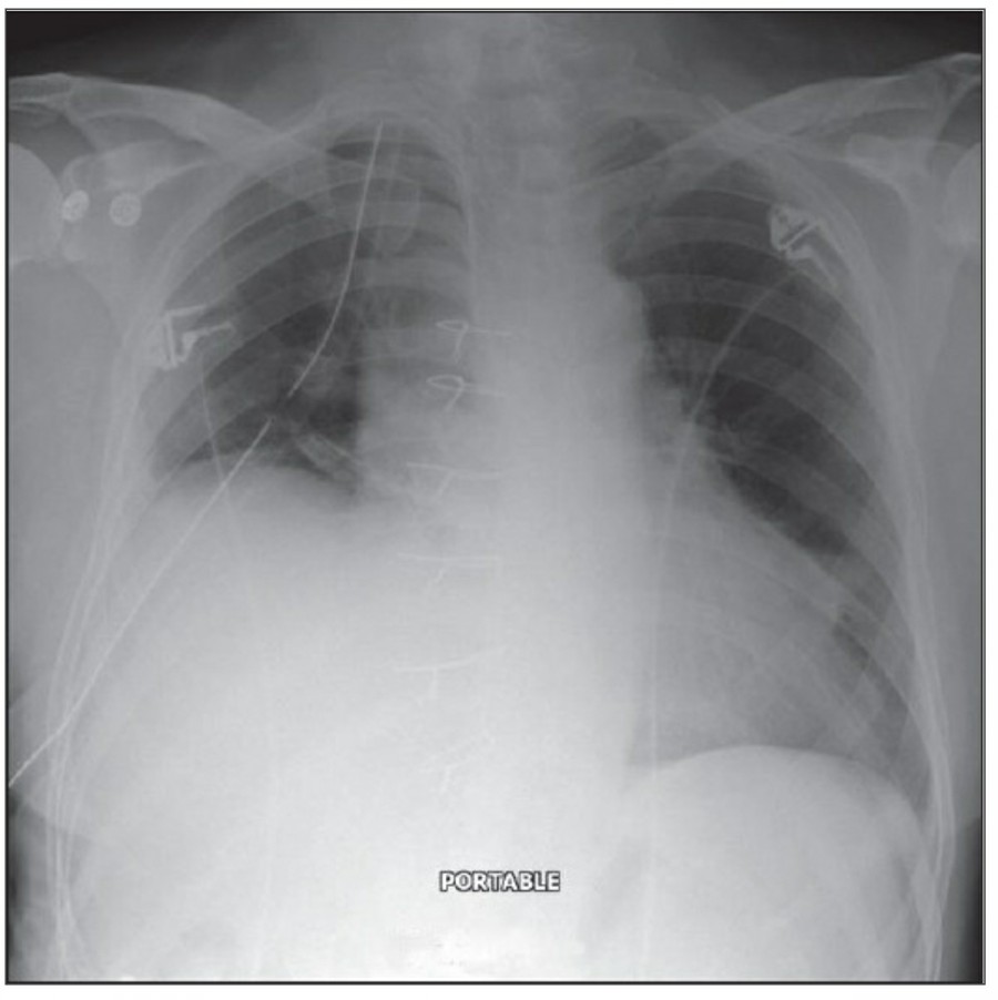
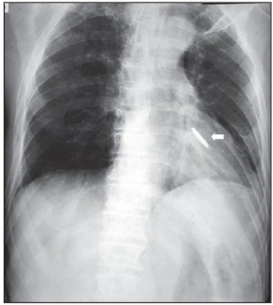
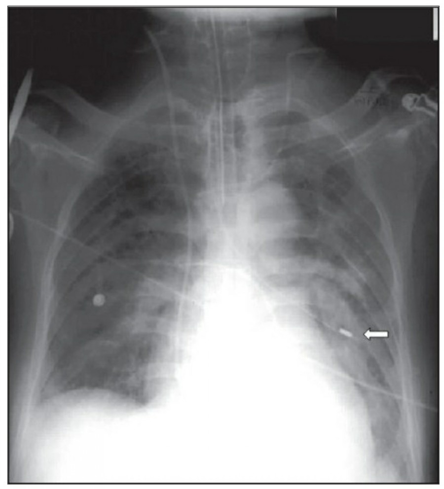
중환자실에서 일반적으로 PA chest X ray는 거의 시행하지 않으며, 주로 AP(anteroposterior) chest X ray를 시행한다. AP CXR은 upright position에서 최대 흡기 시 patient-to-x-ray plate가 72inches(182.88cm)지만, 중환자는 움직임이 제한되므로 supine 혹은 sitting position에서 시행하며 그 거리는 40inches(101.6cm) 정도다.

이렇게 얻어진 image는 gravitational and geometrical effect로 인해 mediastinum과 heart가 확대되어서 보이는데, 더욱이 supine position은 pulmonary vasculature의 physiology를 변화시켜서 혈류가 lung apex로 흐르게 한다-이렇게 얻어진 형태는 PA CXR에서는 비정상으로 간주되지만 AP CXR에서는 정상이다. Supine position은 pleural effuson과 air space shadowing의 감별, pneumothorax 발견을 힘들게 한다.
중환자는 협조가 되지 않거나 수술 후 통증 때문에 full inspiration이 쉽지 않으므로 X ray image를 얻기가 어렵다., 따라서 basialr atelectasis와 pulmonary edema의 진단이 어렵고, heart와 mediastinum의 크기가 정확히 나타나지 않게 된다.

중환자 chest X ray는 민감도와 특이도가 낮다고 하지만, 여러 연구들에서 중환자 chest X ray가 65% 이상의 환자에서 치료 방향에 영향을 줄 수 있는 중요한 병리적 소견을 발견할 수 있다고 서술하고 있다. American College of Radiology에서는 acute cardiopulmonary problem이 있거나 mechanical ventilation 중인 환자에서 daily chest X ray를 촬영할 것을 권고하고 있다. 반면에 cardiac monitoring이 필요하지만 stable 한 환자는 처음 입원 시에만 촬영하는 것으로 충분하며 추가적인 촬영은 indwelling device를 거치하거나 교체한 경우, cardiopulmonary status에 특별한 이상이 의심되는 경우에 할 것을 제안하고 있다.
Henschke CI, Yankelevitz DF, Wand A, Davis SD, Shiau M. Accuracy and efficacy of chest radiography in the intensive care unit. Radiol Clin North Am 1996;34:21-31.
Thoracotomy를 시행받고 나온 환자의 initial postoperative chest X ray에서 각종 line과 endotracheal tube, thoracotomy tube, mediastinal drains, central venous catheter 등을 확인할 수 있을 것이다. 이러한 device들은 그 위치가 제대로 있는지 확인해야 한다.
CABG를 받은 환자에서 lower lobe atelectasis는 흔한데, 주로 왼쪽에 잘 나타나며 수일 내에 별다른 합병증 없이 회복된다. mediastinum도 약간 확대되어 보일 수 있는데, 만약 그 diameter가 많이 증가한다면 mediastinal hemorrhage 등을 시사할 수 있다. CABG 시행 후 약간의 좌측 pleural effusion은 있을 수 있지만 그 양이 많거나 증가한다면 respiratory compromise를 줄이기 위해 intervention이 필요할 수 있다. 따라서 이전의 사진과 비교를 해서 pleural effusion 양의 변화가 있는지 확인이 필요하겠다.

Idendifying lines and tubes and other devices
첫 poratable chest X ray 촬영은 각종 line과 device의 위치를 확인하는 데 꼭 필요하며, 위중한 중환자에서 cardiopulmonary disorder를 evaulation 하기 전에 이 device들 위치에 문제가 있지 않은지 확인해야 한다.


The endotracheal tube
잘못 위치한 entdotracheal tube는 호흡기능에 심각한 영향을 미치는 데 약 10%의 환자에서 보고되고 있다. 따라서 ET tube를 갖고 있는 환자에서는 매일 chest radiographt의 확인이 필요하다. ET tube의 올바른 위치는 mid-trachea lvel에서, carina로부터 약 5cm 위이다. 환자의 고개가 flextion 혹은 extention 하면서 tip의 위치가 바뀔 수 있는데, 가장 최소한의 안전한 위치는 carina로부터 2cm 위이라고 할 수 있다.
Chest X ray 사진에서 carina 위치를 정확히 알 수가 없다면 이전 사진과 비교해서 그 위치를 가늠해 볼 수 있겠지만, upper dorsal spine을 확인해 봄으로써 ET tube의 tip을 확인할 수 있다. Carina는 보통 T4-T5 사이에 위치하므로, ET tube tip이 이 곳에 있다면 제대로 위치하고 있다고 볼 수 있다.


- The Dee method:
Carina의 위치를 확인하는 방법으로서, aortic arch를 먼저 확인한 후 그 중간에서 45' 각도로 inferomedial 하게 내려간다면 midline과 만나는 점에 carina가 위치한다고 볼 수 있다.
Upper airway 손상이 의심될 때, lateral radiograph 촬영이 유용하다. Trachea와 cervical spine 사이 soft tissue space의 지름이 vertebral body 1개의 넓이보다 증가한다면 혈종이나 감염이 원인일 수 있다.
Pneumothorax, pneumomediastinum, subcutaneous emphysema in the neck or precipitous respiratory failure following intubation 등에서는 trachea rupture와 같은 중한 손상을 의심해 볼 수 있는데, trachea rupture는 주로 posterior 쪽에 위치한다.

The thoracostomy tube
Supine AP radiograph에서 air는 anterior에, fluid는 중력에 의해 posterior에 위치하는 걸 인지하고 있어야 한다. Tube가 anterior에 위치하는지 아니면 posterior에 위치하는지는 단 한 장의 AP 사진으로 알기는 어렵다.

Pleural fissure 안에 위치하는 chest tube는 폐 표면이 늘어날 때 종종 배액이 안될 수 있으므로 적절히 기능을 하기 위해서 thoracic cavity 안에 거치되어야 한다. 마지막 side-hole은 radiopaque line의 interruption으로 확인할 수 있는데, 이 지점은 반드시 thoracic cavity 안에 위치해야 한다. Thoracic cavity 밖에 위치하거나 subcutaneous air가 확인되는 경우에 tube가 잘못 들어가 있음을 시사한다. Empyema에서 tube가 제대로 들어가 있지 않다면 배액이 잘 안 되거나 purulent fluid의 loculation이 발생하게 된다.
The feeding tube
Nasogastric tube 삽입은 의식이 없는 환자나 bronchial tree로 들어갈 위험이 높을 때는 chest X ray를 확인해야 한다. 또한 small-bore feeding tube를 삽입하거나 esophagectomy 환자에서 tube를 넣을 때도 chest X ray 확인이 필요하다. NG tube의 lower tip은 보통 abdominal radiograph로서 확인할 수 있다.


Reference
Reading chest radiographs in the critically ill (Part I): Normal chest radiographic appearance, instrumentation and complications from instrumentation. Annals of Thoracic Medicine - Vol 4, Issue 2, April-June 2009
2024.03.01 - [의학] - Reading chest radiographs in the critically ill (Part I) - 1
Reading chest radiographs in the critically ill (Part I) - 1
The Normal Chest X-ray중환자실에서 일반적으로 PA chest X ray는 거의 시행하지 않으며, 주로 AP(anteroposterior) chest X ray를 시행한다. AP CXR은 upright position에서 최대 흡기 시 patient-to-x-ray plate가 72inches(182.88cm
blueorbit.tistory.com
2024.02.08 - [의학] - 중환자에서 chest X ray 촬영 시 고려할 사항
중환자에서 chest X ray 촬영 시 고려할 사항
중환자실에서의 영상 검사 시행은 환자의 거동과 움직임 제한으로 인해 외래 환자와 비교했을 때 고려해야 할 점들이 있으며, 대표적으로 chest X ray를 들 수 있다. AP(anteroposterior) chest X ray 병동과
blueorbit.tistory.com
2023.12.06 - [의학] - 흉부 외상 환자에서 삽입한 chest tube(흉관) 위치의 중요성?
흉부 외상 환자에서 삽입한 chest tube(흉관) 위치의 중요성?
Dose Chest Tube Location Matter? An Analysis of Chest Tube Position and the Need for Secondary Interventions Background 흉부 손상 시 Chest tube(흉관) 삽입은 흔한 치료 방법 중 하나이다. 그동안 chest tube은 특정한 위치로 삽입
blueorbit.tistory.com
'의학' 카테고리의 다른 글
| Reading chest radiographs in the critically ill (Part I)-2 (0) | 2024.03.02 |
|---|---|
| 섬망 발생을 줄이기 위해 멜라토닌을 예방적으로 투여한 효과는? (0) | 2024.03.01 |
| 승압제를 말초정맥으로 투여해도 안전할까? (0) | 2024.02.29 |
| 두부 외상 환자에서 antiplatelet(항혈소판제)와 anticoagulant(항응고제) 치료의 비교 (0) | 2024.02.29 |
| Remdesivir: COVID 19 치료 (0) | 2024.02.29 |


