Parasternal Window
IMAGING WINDOW
전통적으로 cardiac ultrasound exam은 prasternal window에서 시작하는데, 대부분의 환자에서 position에 상관없이 high-quality image를 얻을 수 있는 장점이 있다. Heart가 anterior chest wall과 닿을 수 있도록 환자가 supine 및 left lateral decubitus로 roation 할 수 있다면 가장 좋다. Chronic obstructive pulmonary disease 환자에서는 imaging window를 좀 더 inferior 하게 위치시킴으로써 더 좋은 quality의 image를 얻을 수 있다. Prasternal window는 phased-array transducer를 sternum 바로 좌측의 3rd 혹은 4th intercostal space에 댐으로써 얻을 수 있다. 최적의 window는 2nd와 5th intercostal space 사이 어느 곳에서나 찾을 수 있으므로, provider는 이 부위를 sliding 함으로써 가장 좋은 quality의 image를 얻을 수 있다. 최적의 parastenral window는 ribs과 lung 사이의 interference가 가장 적은 곳에 있는데, 일단 optimal window를 확인한다면, transduce를 sliding 없이 고정하면 된다.
PARASTERNAL LONG-AXIS VIEW
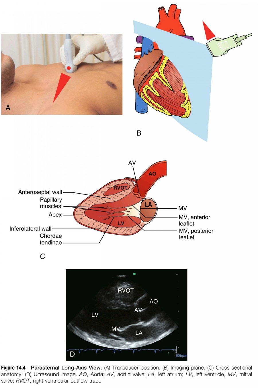
PLAX를 얻기 위해서는 transducer orientation marker를 환자의 right shoulder로 향하게 한다(figure 14.4). Ultrasound beam은 환자의 right shoulder에서 left hip에 이르는 line에 평행하게 위치시킴으로써, cardiac apex에서 base에 이르는 long axis에 해당하는 anatomic cross section의 image를 얻을 수 있다.

Right ventricle(RV)는 screen의 윗 부분에서 anterior에 나타난다. Transducer를 고정한 채 aortic valve와 mitral valve를 확인하면서 ultrasound beam을 LV 상에 위치시킨다. AV와 MV가 같은 plane에 위치하고 ultrasound beam이 LV의 long axis 중간에 위치할 때 최적의 view를 얻을 수 있다. Transducer를 약간 rotation 및 tilting 함으로써 left ventricular cavity가 foreshortening 되는 것을 피하면서 완전히 open 할 수 있다. LV cavity가 축소되게 보이는 error는 흔한데, LV systolic function의 overestimation과 LV cavity dimension을 underestimation 하게 한다. 만약 good-quality image를 얻기가 힘들다면 transducer를 intercostal space를 한 칸 올라가거나 내려가보고 그래도 안되면 다시 처음부터 시도를 해 본다. 아니면, 환자를 left lateral decubitus position으로 변경해 본다. 마지막으로 협조 가능한 환자에게는 의식적으로 respiratory cycle을 바꿔보라고 할 수 있는데, end-expiration에서 호흡을 멈추게 하는 것이 가장 좋다.
PLAX에서 꼭 확인해야 하는 key structure로는 AV, MV, LV, pericardium(heart의 anterior과 posterior 둘 다), right ventricular ounflow tract(RVOT), left ventricular outflow tract(LVOT), portions of the ascending and descending thoracic aorta가 있다. Descending thoracic aorta를 확인하기 위해 depth를 조정해야 한다.
Point-of-care ultrasound 맥락에서 PLAX는 주로 LV size와 function, AV, MV, left atrial size를 평가하는데 이용한다. PLAX는 비록anteroseptal LV wall과 inferolateral LV wall을 보는데 국한되지만, LV systolic function을 정확하게 측정할 수 있다. Pericardial effusion 또한 circumferntial 할 때는 확인 가능하다. RVOT를 작은 cross section으로만 볼 수 있기 때문에 RV size나 function은 제대로 확인할 수 없다; 하지만 severely dilated RV의 발견은 가능하다. PLAX로 AV와 MV의 기본적인 평가와 LVOT level에서 dynamic obstuction의 확인이 가능하다.
PARASTERNAL SHORT-AXIS VIEW
High-quality PSAX view를 빠르게 얻는 가장 좋은 방법은 high-qality PLAX에서 시작하는 것이다. PLAX에서 Transducer를 MV의 가운데에 위치시킨 상태에서 시계 방향으로 90' rotating하면 transducer orientation marker가 환자의 left shoulder를 가리키게 된다(figure 14.5). Chest 위에서 다른 곳으로 sliding 하지 않도록 주의해야 하는데, 두 손을 이용하면 long-axis view에서 short-axis view로 자연스럽게 이동할 수 있다; 한 손은 transducer를 rotation 하고 동시에 다른 손은 transducer를 skin surface에 고정시킨다.

다섯 가지 imaging plane이 Parasternal short-axis view를 얻는 데 이용된다. Point-of-care ultrasound 목적을 고려했을 때, global LV systolic function의 믿을 만한 portroyal을 얻기 위해 midclavicular level이 가장 선호된다. Midclavicular parasternal short-axis view은 cross section에서 양측 papillary muscles이 확인되고 symmetric하게 보일 때 얻을 수 있다. 구형으로 보이는 LV cavity의 cross-sectional image를 얻기 위해서는 transducer를 충분히 rotation 하는 것이 중요한데, oval-shaped LV cavity는 LV function을 잘못 판독할 수 있는 off-axis imaging이나 foreshortening을 보인다.
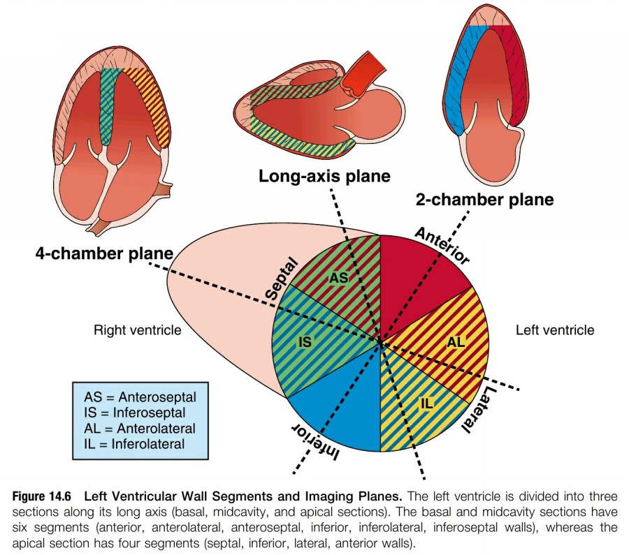
LV systolic function과 segmental LV wall motion을 평가하기 위해서 short-axis midventricular view가 가장 이상적인데, LV wall segments의 nomenclature는 figure 14.6에 있다. 이 view는 RV dilatation과 dysfunction을 평가하는 데 있어 interventricular septum의 shape과 function을 확인하는데 유용하다. Large- 혹은 moderate-sized circumferential pericardial effusion 확인도 가능하다.

Midventricular plane 외 다른 short-axis plane들도 유용한데, figure 14.7은 cardiac base에서 apex에 이르는 순서로 anatomic sequence를 보여준다.

1. Pulmonary artery level:
Midventricular level에서 ultrasound beam을 cardiac base의 superior 쪽으로 tilting한다. Short axis에서 pulmonary valve(PV), main pulmonary artery(MPA), ascending aorta가 확인할 수 있을 때 올바른 plane을 얻을 수 있다. Acute pulmonary embolism(PE)의 rare cases에서 thrombus가 MPA나 proximal left 혹은 right pulmonary artery 안에 있는 것을 발견할 수 있다. Mean pulmonary artery pressure와 diastolic pulmonary pressure를 측정하는 데 pulmonary regurgitation velocity를 이용할 수 있다.
2. AV level:
Pulmonary artery level에서 transduce를 cardiac apex로 향하도록 inferior 쪽으로 약간 tilting한다. Ideal image는 AV의 short-axis view를 포함하는데, AV cusp 세 개 모두 symmetric 하게 나타날 때까지 transducer를 약간 rotating 함으로써 얻을 수 있다. Right atrium(RA), TV, RVOT, left atrium(LA)를 포함하는 ideal image로 AV와 TV의 평가를 할 수 있다.
3. MV level:
Parasternal long-axis view에서 short-axis view로 rotation하면, MV의 독특한 "fish mouth" appearance가 보통 처음으로 나타난다. 이 View로 MV anatomy를 평가할 수 있지만 급성 환자에서는 그 유용성이 제한될 수 있다. MV annulus로 인한 제한 때문에 midventricular level과 비교해서 LV systolic function이 underestimation 될 수 있다.
4. Midventricular, papillary muscle level:
이 view는 급성 환자의 대부분에서 가장 유용한 임상 정보를 제공한다. 양측 papillary muscle이 circular LV cavity의 중간에서 cross section으로 symmetric하게 보인다. 이 level에서 각각의 LV chamber wall segment motion과 전체적인 LV systolic function을 가장 잘 평가할 수 있다.
5. Apical level:
Ultrasound beam을 inferior하게 향하도록 transducer를 tilting 해서 cardiac apex 쪽을 봄으로써 이 short-axis view를 얻을 수 있다. Midpapillary muscle level에서 시작해서 inferior 쪽으로 이동한다면 LV apex를 순차적으로 볼 수 있다. Midventricular level과 비교했을 때 LV systolic function이 overestimation 될 수 있다. Rare case에서 LV apical thrombus를 발견할 수 있다.
Reference
Point-of-Care Ultrasound, 2nd ed. Nilam J. Soni, Robert Arntfield, Pierre Kory. Elsevier
2024.03.06 - [의학] - Cardiac Ultrasound Technique-1
Cardiac Ultrasound Technique-1
Background Point-of-care cardiac ultrasound examination은 experienced provider에게 매우 유용한 임상 도구로서, heart의 structure와 function을 평가해야 하므로 the highest quality image를 얻어야 한다. Cardiac ultrasound exam find
blueorbit.tistory.com
2024.03.07 - [의학] - Cardiac Ultrasound Technique-3
Cardiac Ultrasound Technique-3
Subcostal Window IMAGING WINDOW Point-of-care ultrasound로서 subcostal window는 급성 환자에서 high-yield information을 빠르게 제공한다. Subcostal view는 다음과 같은 장점들이 있다. 1. Subcostal imaging을 위해서 supine positi
blueorbit.tistory.com
2024.02.08 - [의학] - 초음파 유도하 쇄골하 정맥 중심정맥관 삽입술 시 고려할 사항
초음파 유도하 쇄골하 정맥 중심정맥관 삽입술 시 고려할 사항
해부학적 위치 Subclavian vein(쇄골하 정맥)은 axillary vein(액와 정맥)의 근위부이며, 첫 번째 늑골의 lateral edge에서 medial 방향으로 brachiocephalic (innominate) vein(완두 정맥)으로 이어진다. 초음파는 뼈를
blueorbit.tistory.com
'의학' 카테고리의 다른 글
| Cardiac Ultrasound Technique-3 (1) | 2024.03.07 |
|---|---|
| Volume of distribution (0) | 2024.03.07 |
| Cardiac Ultrasound Technique-1 (1) | 2024.03.06 |
| 경장 영양(enteral nutrition) 중인 중환자의 금식 가이드라인 예 (1) | 2024.03.06 |
| ARDS 환자에서 prone position이 venous return에 미치는 영향 (1) | 2024.03.05 |


