Image Acquisition
성인에서 phased-array나 microconvex transducer 등 low-frequency ultrasound transducer로 IVC를 확인할 수 있다. 보통 phased-array transducer를 가장 흔하게 사용하며, 두 가지 technique을 이용한다.
1. Subcostal cardiac window
Subcostal window으로 right atrial junction에서 IVC를 long-axis view로 확인하는 것이 reliability와 reproducibility 면에서 추천되는 방법이다. Phased-array transducer를 subcostal window에 댄 후 transducer orientation marker를 cephalad 쪽으로 향한다(figure 17.4).

Subcostal 4-chamber veiw로 시작해서 RA에 focus를 맞추고 transducer를 반시계방향으로 돌려서 ultrasound beam이 IVC 주행과 일치하게 한다. IVC의 ideal long-axis view는 IVC가 RA로 들어가는 것과 hepatic vein segment가 IVC에 합류하는 것을 보여준다. RA-IVC junction과 hepatic vein(s)를 확인하는 것은 IVC를 옆에 있는 aorta로 잘못 보는 것을 피하게 해 준다. Transducer를 medial 쪽으로 tilting 함으로써 ulsatile, thicker-walled aorta를 확인할 수 있다.
Transducer와 IVC와 longitudinal하게 위치시켜서 IVC를 long axis의 중심에서 보이도록 하는 게 diameter와 collapsibility를 정확하게 측정하는데 중요하다. Off-axis imaging은 vessel를 oblique view로 보이게 해서 small diameter로 오인하게 하는데, 이를 "cylinder effect"라고 한다(figure 17.5). RA-IVC junction과 함께 IVC의 long-axis view가 확보되면, transducer를 tilting 하고 rotating 함으로써 가장 큰 diameter에서 IVC에 beam이 놓이게 된다.

2. Transhepatic coronal view
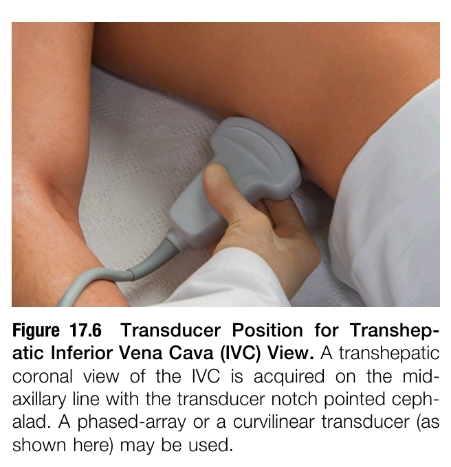
Liver parenchyma는 IVC를 보는데 있어서 매우 뛰어난 acoustic window를 제공하므로, subcostal IVC view를 얻을 수 없을 때(eg, pregnancy, postoperative wounds, dressing, bowel gas, patient discomfort) 유용하다. Transducer를 mid-axillary line에 대고 transducer orientation marker를 cephalad 쪽으로 향한다. Transducer를 posterior 쪽으로 tilting 하면(ie, ultrasound beam을 posterior 쪽으로 향하면) liver와 diaphragm을 지나는 IVC의 long-axis view를 capture 할 수 있다. Aorta는 이 vewi에서 IVC 보다 더 깊은 곳에 위치한다(figure 17.6, 17.7).


IVC diameter와 호흡에 따른 diameter의 변화를 측정하려면, image를 frozen한 후 RA-IVC로부터 2cm 떨어진 지점에서 long axis에 수직으로 caliper를 이용해서 거리를 측정한다. 이 지점이 inter-rater reliability가 좋고 reproducible 하다고 알려져 있다. 가장 큰 diameter를 측정한 후 같은 respiratory cycle 안에서 가장 작은 IVC diameter를 확인하기 위해 cine function을 이용해서 frame에 따라 scroll 한다.
Respiration cycle 동안 IVC diameter의 variation을 확인하기 위해 M-mode를 사용할 수도 있다(figure 17.8). IVC가 증가한 것으로 잘못 판단하는 것을 피하기 위해서 M-mode sampling plane이 IVC에 수직으로 위치하는 것을 확인해야 한다.

Reference
Point-of-Care Ultrasound, 2nd ed. Nilam J. Soni, Robert Arntfield, Pierre Kory. Elsevier
2024.03.06 - [의학] - Cardiac Ultrasound Technique-1
Cardiac Ultrasound Technique-1
Background Point-of-care cardiac ultrasound examination은 experienced provider에게 매우 유용한 임상 도구로서, heart의 structure와 function을 평가해야 하므로 the highest quality image를 얻어야 한다. Cardiac ultrasound exam find
blueorbit.tistory.com
'의학' 카테고리의 다른 글
| 중환자에서 발생하는 의식 변화 (0) | 2024.03.09 |
|---|---|
| COVID-19 환자에서 interlekin-6, C-reactive protein, procalcitonin와 예후의 관계는? (0) | 2024.03.09 |
| 중환자에서 glutamine의 공급 (0) | 2024.03.08 |
| 초음파를 이용한 interior vena cava(하대정맥)의 평가_1 (1) | 2024.03.08 |
| Cardiac Ultrasound Technique-3 (1) | 2024.03.07 |


